Fasciculus gracilis

Fasciculus gracilis
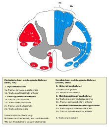
Querschnitt durch das Rückenmark, 3a bezeichnet den Fasciculus gracilis

Der Fasciculus gracilis ist eine Nervenbahn innerhalb des Rückenmarks. Er leitet epikritische und propriozeptive Informationen, vor allem von der unteren Extremität zum Gehirn. Der Fasciculus gracilis gehört mit dem Fasciculus cuneatus zu den Hinterstrangbahnen des Rückenmarks.

Die Nervenfasern des Fasciculus gracilis verlaufen gleichseitig (ipsilateral) ohne vorherige Umschaltung zum Nucleus gracilis in der Medulla oblongata, wo sie auf das zweite Neuron umgeschaltet werden. Nach dieser Umschaltung setzen sie sich als Lemniscus medialis fort, in dem die Kreuzung auf die Gegenseite (kontralateral) erfolgt.

Der Fasciculus gracilis wird auch als Goll-Bündel bezeichnet. Der Name geht auf die Erstbeschreibung der anatomischen Struktur aus dem Jahr 1860 durch den schweizerischen Neuroanatom Friedrich Goll zurück.[1]

Literatur

  • Martin Trepel: Neuroanatomie mit Studentconsultzugang: Struktur und Funktion. Elsevier,Urban&FischerVerlag, 4. Auflage 2008, ISBN 9783437412981, S. 106–107.

Einzelnachweise

  1. Mumenthaler M.: Zur Geschichte der Schweizerischen Neurologischen Gesellschaft [Referat]. Schweiz Arch Neurol Psychiatr. 2000; 151:168–72. PDF-Version

Wikimedia Foundation.

Игры ⚽ Нужна курсовая?

Schlagen Sie auch in anderen Wörterbüchern nach:

  • Fasciculus gracilis — Infobox Anatomy Name = PAGENAME Latin = f. gracilis medullae spinalis GraySubject = 185 GrayPage = 762 Caption = Fasciculus cuneatus is 3a, in blue at upper right. Caption2 = Diagram of the principal fasciculi of the spinal cord. Width = 325… …   Wikipedia

  • Fasciculus gracilis — grakštusis pluoštelis statusas T sritis centrinė nervų sistema atitikmenys: lot. Fasciculus gracilis ryšiai: platesnis terminas – viršutinis pluoštas …   Paukščių anatomijos terminai

  • Fasciculus gracilis — grakštusis pluoštelis statusas T sritis gyvūnų anatomija, gyvūnų morfologija atitikmenys: lot. Fasciculus gracilis ryšiai: platesnis terminas – dorsalinis pluoštas …   Veterinarinės anatomijos, histologijos ir embriologijos terminai

  • Fasciculus gracilis — grakštusis pluoštelis statusas T sritis gyvūnų anatomija, gyvūnų morfologija atitikmenys: lot. Fasciculus gracilis ryšiai: platesnis terminas – pailgosios smegenys …   Veterinarinės anatomijos, histologijos ir embriologijos terminai

  • Fasciculus gracilis — grakštusis pluoštelis statusas T sritis gyvūnų anatomija, gyvūnų morfologija atitikmenys: lot. Fasciculus gracilis ryšiai: platesnis terminas – pailgųjų smegenų pjūviai …   Veterinarinės anatomijos, histologijos ir embriologijos terminai

  • fasciculus gracilis — fasciculus grac·i·lis gras ə ləs n either of a pair of nerve tracts of the posterior funiculus of the spinal cord situated on opposite sides of and immediately adjacent to the posterior median septum and carrying nerve fibers from the lower part… …   Medical dictionary

  • fasciculus gracilis medullae oblongatae — [TA] the continuation into the medulla oblongata of the fasciculus gracilis of the spinal cord; called also posteromedian column of medulla oblongata …   Medical dictionary

  • fasciculus gracilis medullae spinalis — [TA] the median portion of the posterior funiculus of the spinal cord, composed of ascending fibers that terminate in the nucleus gracilis of the medulla oblongata; called also Goll column …   Medical dictionary

  • Fasciculus gracilis — (funiculus) тонкий пучок, или пучок Gall я вместе с клиновидным пучком составляет так назыв. задний столб спинного мозга и лежит между указанным пучком и septum medianum posterius (задней средней перегородкой) …   Энциклопедический словарь Ф.А. Брокгауза и И.А. Ефрона

  • fasciculus cuneatus — fasciculus cu·ne·atus .kyü nē āt əs n either of a pair of nerve tracts of the posterior funiculus of the spinal cord that are situated on opposite sides of the posterior median septum lateral to the fasciculus gracilis and that carry nerve fibers …   Medical dictionary

Share the article and excerpts

Direct link
Do a right-click on the link above
and select “Copy Link”