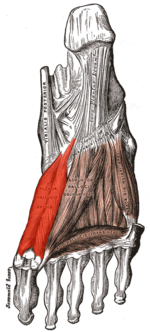
Musculus flexor hallucis brevis
- Musculus flexor hallucis brevis
-
| Musculus flexor hallucis brevis |
 |
| Fußsohlenmuskulatur des Menschen |
| Ursprung |
| v. a. Os cuneiforme intermedium |
| Ansatz |
| Grundglied der großen Zehe (beide Köpfe) und mediales Sesambein (für den medialen Kopf) bzw. laterales Sesambein (für den lateralen Kopf) der Großzehe |
| Funktion |
| Beugung (Plantarflexion) der Großzehe |
| Innervation |
| Äste des Nervus tibialis: N. plantaris medialis (für den medialen Kopf) und N. plantaris lateralis (für den lateralen Kopf) |
| Spinale Segmente |
| S1, S2 |
Der Musculus flexor hallucis brevis (lat.: Kurzer Großzehenbeuger) ist ein Skelettmuskel im Bereich der Unterseite des Fußskelettes. Er unterteilt sich in einen lateralen (äußeren) und einen medialen (inneren) Muskelbauch.
Ursprung
Der Ursprung liegt im Wesentlichen im Bereich des Mittleren Keilbeins (Os cuneiforme intermedium). Darüber hinaus entspringt der Muskel aber auch den folgenden Strukturen:
- Inneres Keilbein (Os cuneiforme mediale)
- Äußeres Keilbein (Os cuneiforme laterale)
- Langes Sohlenband (Ligamentum plantare longum)
- Ligamentum calcaneocuboideum plantare
- Sehne des hinteren Schienbeinmuskels (Musculus tibialis posterior)
Ansatz
Der Musculus flexor hallucis brevis unterteilt sich in einen lateralen und einen medialen Muskelbauch:
- Der laterale Anteil setzt am lateralen Sesambein der Kapsel des Großzehengrundgelenkes und der Basis des Grundgliedes der Großzehe an.
- Der mediale Anteil setzt am medialen Sesambein der Kapsel des Großzehengrundgelenkes und der Basis des Grundgliedes der Großzehe an.
Funktion
Der Musculus flexor hallucis brevis beugt die große Zehe im Großzehengrundgelenk (körpernahes Gelenk).
Siehe auch
Kategorie: - Skelettmuskel der unteren Extremität
Wikimedia Foundation.
Schlagen Sie auch in anderen Wörterbüchern nach:
Musculus flexor hallucis brevis — trumpasis tiesiamasis pirmojo piršto raumuo statusas T sritis raumenys atitikmenys: lot. Musculus flexor hallucis brevis ryšiai: platesnis terminas – kojos raumenys … Paukščių anatomijos terminai
flexor hallucis brevis muscle — musculus flexor hallucis brevis … Medical dictionary
musculus flexor hallucis brevis — [TA] short flexor muscle of great toe: origin, undersurface of cuboid, lateral cuneiform; insertion (2 heads): LATERAL HEAD—lateral side of base of proximal phalanx of toe; MEDIAL HEAD—medial side of base of proximal phalanx of toe;… … Medical dictionary
Flexor hallucis brevis — Musculus flexor hallucis brevis Fußsohlenmuskulatur des Menschen Ursprung v. a. Os cuneiforme intermedium Ansatz Grundglied der großen Zehe … Deutsch Wikipedia
Flexor hallucis brevis muscle — Muscle infobox Name = Flexor hallucis brevis muscle Latin = musculus flexor hallucis brevis GraySubject = 131 GrayPage = 493 Caption = Muscles of the sole of the foot. Third layer. (Flexor hallucis brevis visible at left.) Origin = Insertion =… … Wikipedia
M. flexor hallucis brevis — Musculus flexor hallucis brevis Fußsohlenmuskulatur des Menschen Ursprung v. a. Os cuneiforme intermedium Ansatz Grundglied der großen Zehe … Deutsch Wikipedia
flexor muscle of great toe short — musculus flexor hallucis brevis … Medical dictionary
Extensor hallucis brevis muscle — Muscle infobox Name = Extensor hallucis brevis Latin = musculus extensor hallucis brevis GraySubject = 129 GrayPage = 490 Caption = The mucous sheaths of the tendons around the ankle. Medial aspect. (Ext. hall. long. labeled at top center.)… … Wikipedia
musculus — SYN: muscle.For histologic description, see muscle. [L. a little mouse, a muscle, fr. mus (mur ), a mouse] musculi abdominis SYN: muscles of abdomen, under muscle. m. abductor [TA] SYN: abductor (muscle). m. abductor … Medical dictionary
Músculo flexor corto del dedo gordo — Flexor corto del dedo gordo Vista de la planta del pie humano en la que puede observarse en color rojo brillante el músculo. Latín Musculus flexor hallucis … Wikipedia Español

