- Myeloblastin
-
Proteinase 3 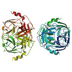
nach PDB 1FUJ Vorhandene Strukturdaten: 1fuj Größe 221 aa; 24,2 kDa Bezeichner Gen-Namen PRTN3; PR3; ACPA; AGP7; C-ANCA; MBT; P29; PR-3 Externe IDs OMIM: 177020 UniProt: P24158 MGI: 893580 Enzymklassifikation EC, Kategorie 3.4.21.76 Serinprotease MEROPS S01.134 Reaktionsart Hydrolyse von Proteinen incl. Elastin Substrat Protein mit Aminosäure -Ala-+-Xaa- oder -Val-+-Xaa- Produkte Spaltprodukte Vorkommen Homologie-Familie Trypsin Precursor Übergeordnetes Taxon Lebewesen Orthologe Mensch Maus Entrez 5657 19152 Ensembl ENSG00000196415 ENSMUSG00000057729 UniProt P24158 Q61096 Refseq (mRNA) NM_002777 NM_011178 Refseq (Protein) NP_002768 NP_035308 Genlocus Chr 19: 0.79 - 0.8 Mb Chr 10: 79.28 - 79.29 Mb PubMed Suche [1] [2] Proteinase 3 (PR3, Myeloblastin) ist ein Enzym in Fischen und Vierbeinern (Euteleostomi), das mit seiner antimikrobiellen Wirkung eine Rolle im Immunsystem spielt. Es handelt sich um eine Serinprotease, die mit der neutrophilen Elastase (ELA2) und Azurocidin (AZU) verwandt ist, auf demselben Genabschnitt kodiert ist und mit ihnen zusammen in neutrophile Granulozyten eingebaut wird. Bei einer Autoimmunerkrankung im Menschen, der Wegener-Granulomatose, werden Antikörper gegen das Enzym gebildet.[1]
Synthese
Das PR3-Gen auf Chromosom 19 ist über 6,5 Kilobasen und 5 Exons verteilt. Das Transkript hat eine Länge von 1001 Basen und kodiert für 256 Aminosäuren, welche nach Spleißen der Signalsequenz und des Propeptids ein Protein mit 221 Aminosäuren und 24 kDa Molmasse ergeben. Durch N-Glykosilierung an den Aminosäuren 129 und 174 (gerechnet vom ungespleißten Protein) entsteht schließlich die 29 kDa schwere Proteinase 3, ein kationisches Glykoprotein.[2][3]
Biologische Funktion
Proteinase 3 ist Bestandteil der primären (azurophilen, α-) Granula der neutrophilen Granulozyten und Lysosomen der Monozyten. Das Enzym kann bereits in frühen Phasen der Entwicklung von Monzyten und Granulozyten nachgewiesen werden. Werden Granulozyten aktiviert, verschmelzen die α-Granula mit der Zellmembran und setzen so Proteinase 3 frei. In vitro kann dieser Effekt durch TNF-α ausgelöst werden. Proteinase 3 hat proteolytische Eigenschaften und spaltet z. B. Elastin, Fibronectin, Laminin, Hämoglobin und Kollagen Typ IV. Proteinase 3 wirkt antimikrobiell gegen E. coli und C. albicans und ist an der Reifung myeloider Zellen beteiligt. Der natürliche Inhibitor von Proteinase 3 ist α-1-Antitrypsin.[4][5][6][7][8][9][10][11][12]
Einzelnachweise
- Lothar Thomas: Labor und Diagnose 5. Auflage, Frankfurt/Main 2000, ISBN 3980521559
- ↑ OMIM-Eintrag
- ↑ Ensembl-Eintrag
- ↑ UniProt-Eintrag
- ↑ von Vietinghoff S, Eulenberg C, Wellner M, Luft FC, Kettritz R: Neutrophil surface presentation of the anti-neutrophil cytoplasmic antibody-antigen proteinase 3 depends on N-terminal processing. In: Clin. Exp. Immunol.. 152, Nr. 3, June 2008, S. 508–16. doi:10.1111/j.1365-2249.2008.03663.x. PMID 18462208
- ↑ Peikert T, Finkielman JD, Hummel AM, et al: Functional characterization of antineutrophil cytoplasmic antibodies in patients with cocaine-induced midline destructive lesions. In: Arthritis Rheum.. 58, Nr. 5, May 2008, S. 1546–51. doi:10.1002/art.23469. PMID 18438818
- ↑ Kallenberg CG: Pathogenesis of PR3-ANCA associated vasculitis. In: J. Autoimmun.. 30, Nr. 1-2, 2008, S. 29–36. doi:10.1016/j.jaut.2007.11.005. PMID 18162369
- ↑ Hajjar E, Mihajlovic M, Witko-Sarsat V, Lazaridis T, Reuter N: Computational prediction of the binding site of proteinase 3 to the plasma membrane. In: Proteins. 71, Nr. 4, June 2008, S. 1655–69. doi:10.1002/prot.21853. PMID 18076025
- ↑ Korkmaz B, Moreau T, Gauthier F: Neutrophil elastase, proteinase 3 and cathepsin G: physicochemical properties, activity and physiopathological functions. In: Biochimie. 90, Nr. 2, February 2008, S. 227–42. doi:10.1016/j.biochi.2007.10.009. PMID 18021746
- ↑ Müller A, Voswinkel J, Gottschlich S, Csernok E: Human proteinase 3 (PR3) and its binding molecules: implications for inflammatory and PR3-related autoimmune responses. In: Ann. N. Y. Acad. Sci.. 1109, August 2007, S. 84–92. doi:10.1196/annals.1398.010. PMID 17785293
- ↑ Sugawara S: Immune functions of proteinase 3. In: Crit. Rev. Immunol.. 25, Nr. 5, 2005, S. 343–60. PMID 16167885
- ↑ Wu YY, Hsu TC, Chen TY, et al: Proteinase 3 and dihydrolipoamide dehydrogenase (E3) are major autoantigens in hepatitis C virus (HCV) infection. In: Clin. Exp. Immunol.. 128, Nr. 2, May 2002, S. 347–52. PMID 11985526
- ↑ He Y, Young PK, Grinnell F: Identification of proteinase 3 as the major caseinolytic activity in acute human wound fluid. In: J. Invest. Dermatol.. 110, Nr. 1, January 1998, S. 67–71. doi:10.1046/j.1523-1747.1998.00075.x. PMID 9424090

Wikimedia Foundation.